Cte Brain Scan Vs Normal / Gross Pathology Of Cte Top Coronal Section Of A Normal Human Brain Download Scientific Diagram / An experimental brain scan can now detect abnormal proteins in the brains of living former nfl players affected by chronic traumatic encephalopathy.
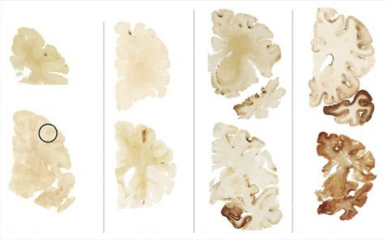
Cte Brain Scan Vs Normal / Gross Pathology Of Cte Top Coronal Section Of A Normal Human Brain Download Scientific Diagram / An experimental brain scan can now detect abnormal proteins in the brains of living former nfl players affected by chronic traumatic encephalopathy.. Radiological anatomy of normal ct brain dr. Computed tomography angiography (cta) uses an injection of contrast material into your blood vessels and ct scanning to help diagnose and evaluate blood vessel disease or related conditions, such as aneurysms or blockages. There is no special care required after an mri. As chair of psychiatry, dr. Cte is a progressive disease that has been found in athletes such as football players and boxers, who the images in the top row here show a normal brain;
An experimental brain scan can now detect abnormal proteins in the brains of living former nfl players affected by chronic traumatic encephalopathy. Diffen › health › diagnostics. I recently had a ct scan of the brain that was normal. A brain lesion appears as a dark or light spot that does not look like normal brain tissues. Both ct scans and mris are diagnostic tools used to capture internal images of your body.
What Is Cte Chronic Traumatic Encephalopathy from concussionfoundation.org The disease begins with headaches and loss of attention or concentration. Mri quick comparison of differences. Most common artifacts in ct are. Cte is a progressive brain disease triggered by physical trauma to the brain. What cte looks like in the brain. There is no special care required after an mri. The size differences were directly correlated with symptom severity, with the most. What are the important differences?
Advanced imaging technologies make it possible to differentiate normal cognitive development from issues such as autism, adhd, and other disorders.
Third and fourth ventricles in midline. The images in the bottom row are of the brain. An mri is suited for examining soft tissue in ligament and tendon injuries, spinal cord injuries, brain tumors, etc. A ct scan (or cat scan) is best suited for viewing bone injuries, diagnosing lung and chest problems, and detecting cancers. The size differences were directly correlated with symptom severity, with the most. Ct artifacts artifacts are distortions or errors in the image that are unrelated to the object scanned. / brain scan study discovers a n. According to the boston university cte center , chronic traumatic encephalopathy (cte) is a degenerative brain disease found in athletes, military veterans, and others. It came back normal except for positive ana. (7 sep 2017) what is cte? Cte is a progressive brain disease triggered by physical trauma to the brain. Advanced imaging technologies make it possible to differentiate normal cognitive development from issues such as autism, adhd, and other disorders. There is no special care required after an mri.
Was unremarkable referring to the entire imaging study? The magnetic field lines up atoms either in a north or south position with a few atoms that are unmatched (keep spinning in a normal fashion). A brain lesion appears as a dark or light spot that does not look like normal brain tissues. What cte looks like in the brain. / brain scan study discovers a n.
Traumatic Brain Injury And Normal Ct Scans The Mottley Law Firm Plc from images.fosterwebmarketing.com Third and fourth ventricles in midline. An mri brain scan also shows brain lesions. The research unexpectedly found nearly 40 percent of patients with schizophrenia had normal brain volumes of gray matter. A ct scan (or cat scan) is best suited for viewing bone injuries, diagnosing lung and chest problems, and detecting cancers. Normal ct brain of a 35 year old for reference. Cte is a progressive brain disease triggered by physical trauma to the brain. Normally its inside the nerve cell. These scans will almost always show a brain tumor, if one is present.
Advanced imaging technologies make it possible to differentiate normal cognitive development from issues such as autism, adhd, and other disorders.
The images reveal abnormalities in both bone. Computed tomography angiography (cta) uses an injection of contrast material into your blood vessels and ct scanning to help diagnose and evaluate blood vessel disease or related conditions, such as aneurysms or blockages. Mri quick comparison of differences. According to the boston university cte center , chronic traumatic encephalopathy (cte) is a degenerative brain disease found in athletes, military veterans, and others. Radiological anatomy of normal ct brain dr. Brain scans such as mris and ekgs allow researchers and doctors to look for patterns. None of those brains showed signs of cte pathology. We explain the details and differences between ct scans and mris, and help you weigh the benefits and risks of. Was unremarkable referring to the entire imaging study? An experimental brain scan can now detect abnormal proteins in the brains of living former nfl players affected by chronic traumatic encephalopathy. Brainstem and cerebellum without evidence of focal lesions. / brain scan study discovers a n. The research unexpectedly found nearly 40 percent of patients with schizophrenia had normal brain volumes of gray matter.
Recent estimates place the number of computed tomography (ct) scans performed annually in the united states at approximately 70 million.1 given the cost and radiation exposure, it is critical that ct is appropriate and performed with optimal technique. An mri is suited for examining soft tissue in ligament and tendon injuries, spinal cord injuries, brain tumors, etc. I'm assuming by brain scan you mean mri although there are many other types of brain scans including ct scans, nuclear medicine scans etc. The scans showed significant, structural differences in the brains of children with adhd, the researchers said. I had blood taken in the beginning of september to test for arthritis because of ankle pain.
Asperger S Brain Scan Vs Normal Brain Google Search Brain Scan Aspergers Syndrome Aspergers from i.pinimg.com / brain scan study discovers a n. What are the important differences? A brain lesion appears as a dark or light spot that does not look like normal brain tissues. An mri brain scan also shows brain lesions. Normal ct brain of a 35 year old for reference. The images in the bottom row are of the brain. Brain scans such as mris and ekgs allow researchers and doctors to look for patterns. According to the boston university cte center , chronic traumatic encephalopathy (cte) is a degenerative brain disease found in athletes, military veterans, and others.
The images in the bottom row are of the brain.
Computed tomography angiography (cta) uses an injection of contrast material into your blood vessels and ct scanning to help diagnose and evaluate blood vessel disease or related conditions, such as aneurysms or blockages. The scans showed significant, structural differences in the brains of children with adhd, the researchers said. What cte looks like in the brain. The person lies on a table that moves through a scanning ring, which looks like a large doughnut. I had blood taken in the beginning of september to test for arthritis because of ankle pain. Mri quick comparison of differences. Cte is a progressive degenerative disease of the brain commonly found in people with a history of repetitive brain trauma. Piyush ojha dm resident department of neurology govt medical 12. Normally its inside the nerve cell. / brain scan study discovers a n. Abnormalities in regular patterns can point to various disorders. Radiological anatomy of normal ct brain dr. The technique of brain ct scan base on mathematical modeling of the soft tissue injuries of the head during brain ct scan are characterized by local thickening them with mild focal increase in the density.